
Case Reports in Clinical Cardiology Journal | Cambridge City
  • info.clinical@cardiologycasereportsjournal.org
  • CB1 OBG, Cambridge City, UK
  • Submit Manuscript

Article Details

Case Report
Volume 6, Issue 3

Transesophageal Echocardiography-Guided Management of Acute Left Main Coronary Thromboembolism During Redo Aortic Valve Surgery

Rézan Watti1*, Győző Wrana2, Károly Gombocz2, Jezdancher Watti3 and Aref Rashed1

1Department of Cardiac Surgery, Zala County St. Rafael Hospital, Zalaegerszeg, Hungary
2Department of Cardiac Surgery Intensive Care Unit, Zala County St. Rafael Hospital, Zalaegerszeg, Hungary
3Department of Internal Medicine, Bugát Pál Hospital, Gyöngyös, Hungary

*Corresponding author: Rézan Watti, Department of Cardiac Surgery, Zala County St. Rafael Hospital, Zalaegerszeg, Hungary.
E-mail: kurdishrezan1@gmail.com

Received: May 03, 2026; Accepted: May 21, 2026; Published: June 05, 2026

Citation: Watti R, Wrana G, Gombocz K, et al. Transesophageal Echocardiography-Guided Management of Acute Left Main Coronary Thromboembolism During Redo Aortic Valve Surgery. Case Rep Clin Cardiol J. 2026; 6(3): 182.
Transesophageal Echocardiography-Guided Management of Acute Left Main Coronary Thromboembolism During Redo Aortic Valve Surgery
Abstract

Background: Redo aortic valve surgery is associated with high perioperative risk, and intraoperative hemodynamic instability can be life-threatening. Acute left main coronary thromboembolism is an exceptionally rare complication that requires immediate diagnosis and intervention. Transesophageal echocardiography provides real-time diagnostic capability in the operating room.

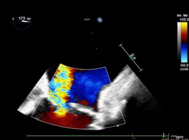
Methods: We present the case of a 31-year-old patient who underwent redo aortic valve replacement for prosthetic valve thrombosis. During weaning from cardiopulmonary bypass, the patient developed severe left ventricular dysfunction associated with regional wall motion abnormalities. Real-time transesophageal echocardiography identified a newly developed flow obstruction in the left main coronary artery, prompting urgent surgical reintervention.

Results: A fresh thrombus was extracted from the left main coronary artery using a Fogarty catheter under transesophageal echocardiography guidance. Intraoperative echocardiography confirmed restoration of coronary flow and rapid improvement in ventricular function, with the ejection fraction increasing from 25% to 45%. The patient was successfully weaned from extracorporeal support and had an uneventful postoperative recovery.

Conclusion: This case highlights the critical importance of structured intraoperative diagnostics and the pivotal role of TEE in the rapid identification and management of rare, life-threatening complications during complex cardiac surgery. Transesophageal echocardiography was instrumental in guiding both diagnosis and immediate surgical intervention, resulting in a favorable outcome.

Keywords: Redo aortic valve surgery; Left main coronary thromboembolism; Transesophageal echocardiography; Intraoperative diagnosis; Real-time imaging; Cardiac surgery complications; Fogarty catheter; Perioperative imaging