Case Reports in Clinical Cardiology Journal | Cambridge City
  • info.clinical@cardiologycasereportsjournal.org
  • CB1 OBG, Cambridge City, UK
  • Submit Manuscript

Article Details

Clinical Image
Volume 2, Issue 2

Calcified Aorta Urine Lithiasis and Hydroxyapatite Stars

Francisco-Javier Roldan-Gómez1, Gabriela Rodríguez-Echeverría1, Carlos Linares Lopez2 and Aurora de la Peña-Díaz1,2*

1Outpatient and Molecular Biology Departments at the Instituto Nacional de Cardiología Ignacio Chávez (INC), Mexico City, Mexico

2Medicine Faculty, pharmacology Department and Geophysics Institute at the Universidad Nacional Autónoma de México (UNAM), Mexico City, Mexico

*Corresponding author: Aurora de la Peña Díaz, Instituto Nacional de Cardiología Ignacio Chávez, Juan Badiano Núm, 1 Col, Belisario Domínguez Sección XVI, Tlalpan, CP 14080, Mexico City, Mexico.

Received: October 21, 2022; Accepted: November 02, 2022; Published: November 15, 2022

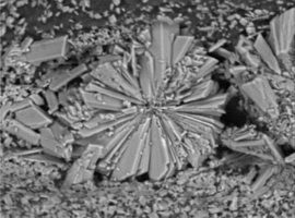
Citation: Roldan-Gómez FJ, Rodríguez-Echeverría G, Lopez CL, Peña-Díaz ADL. Calcified Aorta, Urine Lithiasis and Hydroxyapatite Stars. Case Rep Clin Cardiol J. 2022; 2(2): 114.

Calcified Aorta Urine Lithiasis and Hydroxyapatite Stars
Abstract

Calcification is a key process in the development of degenerative aortic stenosis. Spherical crystalline particles, composed of calcium (Ca2+) and phosphorus (PO43-) and, in smaller portions, a compacted material of magnesium (Mg), carbon (C) and fibers are the starting point of the aortic calcification process. Hydroxyapatite (HA) is a very versatile mineral formed by calcium phosphate in amorphous (the most frequent) or crystalline structures. The morphology of HA crystals depends on Ca/P molar ratios, temperature, pH, movement and pressure. In artificial conditions, we are able to obtain HA crystallization in acicular shapes for medical applications (bone, dental and filler material). Changing environment variables, we can get also long plate-like crystals, hexagonal prism-like crystals, and flakes forms. Finding star-shaped HA crystals in nature is uncommon. In humans, we had only observed this form in urine and now, by of SEM (scanning electron microscopy), in the aortic valve of patients with degenerative stenosis. As crystal shape depends on environmental conditions, knowing the shared factors that lead to certain morphologies could us better understand mechanisms of calcification in biological tissues.