Case Reports in Clinical Cardiology Journal | Cambridge City
  • info.clinical@cardiologycasereportsjournal.org
  • CB1 OBG, Cambridge City, UK
  • Submit Manuscript

Article Details

Case Report
Volume 4, Issue 1

Adult T-type Lymphoblastic Lymphoma Presenting as Inferior Wall Myocardial Infarction: A Case Report

Abdelrahman Elhakim1*, Mohamed Gayed2, Mohamed Elhakim3 and Mohamed Saad4

1Interventional Cardiology Consultant, Schoen Hospital Neustadt, Am Kiebitzberg 10, Neustadt in Holstein, Germany
2Interventional Cardiologist, Medinos Hospital Sonneberg, Neustadter Street 61, Sonneberg, Germany
3Critical Care and Anesthesia Medical Officer, Nepean Hospital, Derby Street, Kingswood NSW, Sydney, Australia
4Interventional Cardiology Consultant, Schleswig-Holstein University Hospital-Kiel, Arnold-Heller-Street 3, Kiel, Germany

*Corresponding author: Abdelrahman Elhakim, Interventional Cardiology Consultant, Schoen Hospital Neustadt, AM Kiebitzberg 10, 23730, Neustadt in Holstein, Germany.

Received: May 14, 2023; Accepted: May 26, 2023; Published: July 05, 2023

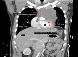
Citation: Elhakim A, Gayed M, Elhakim M, Saad M. Adult T-type Lymphoblastic Lymphoma Presenting as Inferior Wall Myocardial Infarction: A Case Report. Case Rep Clin Cardiol J. 2023; 3(3): 131.

Adult T-type Lymphoblastic Lymphoma Presenting as Inferior Wall Myocardial Infarction: A Case Report
Abstract

Background: Lymphoblastic lymphoma (LBL) is the second most common type of non-Hodgkin lymphoma (NHL) in childhood and adolescence, accounting for 25–35% of all cases. The majority, 70–80%, is of T-lymphoblastic origin, while 20–25% arises from B lymphoblasts. It is characterized by a large mediastinal mass with less than 25% bone marrow involvement. Most patients present with a cough, dyspnea, or chest pain, which is incidentally detected by a chest X-ray. Extracardiac compression of cardiac and mediastinal structures can occur, causing variable clinical presentation according to a structure being compressed, such as myocardial infarction.

Case presentation: We report a case of T-cell lymphoma, which was initially suspected by a chest X-ray and CTA, in a 31-year-old male who presented in the emergency room for severe chest pain and had a history of smoking. An ECG revealed a STEMI of the inferior myocardial wall. Transthoracic echocardiography showed circumferential pericardial effusion of 5mm, and CT aortography revealed an extensive and dense large soft tissue mass (measuring 85 x 105 x 115mm), which had contact with the coronary LAD artery. Coronary angiography revealed medial LAD stenosis of about 50% stenosis, which was mostly due to compression outside the vessel wall. A thymic carcinoma was confirmed by biopsy and the patient received radiotherapy (GMALL protocol). The further evaluation and follow up for 90 days was uneventful.

Conclusion: T-type lymphoblastic lymphoma can present as inferior wall myocardial infarction. Extra care should be taken on extra cardiac causes of myocardial infarction.

Keywords: Chest pain; Mediastinal mass; Thymic carcinoma, Lymphoblastic lymphoma; Case report